Long Bone Diagram Endosteum : Bone Development And Growth Intechopen / Both the periosteum and the.. At the ends of the bone the periosteum is continuous with the joint. The bones in your body have 3 major types of bone cells. The endosteum is located on the internal surface of the bone, being the membranous layer that covers the medullary cavity, the bony trabeculae (spongy part of the bone), the haversian canals and internal walls of the compact long bones. Make sure that you follow all the guidelines for biological drawings: Figure 6.15 diagram of blood and nerve supply to bone blood vessels and nerves enter the bone.
Its not option b because a fossa is a animal that is in the cat species. It is important to note that the absence of endosteum or periosteum on a bone signals that the bone is ready to be reabsorbed by osteoclasts. Learn about long bone diagram with free interactive flashcards. Long bones, especially the femur and tibia, are subjected to most of the load during daily activities and they are crucial for skeletal mobility. Let's start by looking at a diagram of bone tissue.
Bone Terminology Diagram Radiology Case Radiopaedia Org from prod-images-static.radiopaedia.org The thigh bone (femur) is a long bone. The diaphysis and the epiphysis. This endosteal surface is usually resorbed during long periods of malnutrition, resulting in less cortical thickness. This page is about endosteum of bone,contains anatomy of a long bone ms. The endosteum can be seen in the t.s. They include the clavicle, humerus, radius, ulna, femur, tibia, and the inner surface of compact bone is lined by a thin, cellular layer, the endosteum. The long bones' tubular design provides maximum strength with minimum weight. Blood vessels and tissue surrounding the injured area bleed and if medullary lesions develop along the inner aspect of the cortical bones, especially in the long bones, endosteal scalloping may be observed.
Bone marrow is found in the bone cavities of long bones and is involved in the production of blood cells.
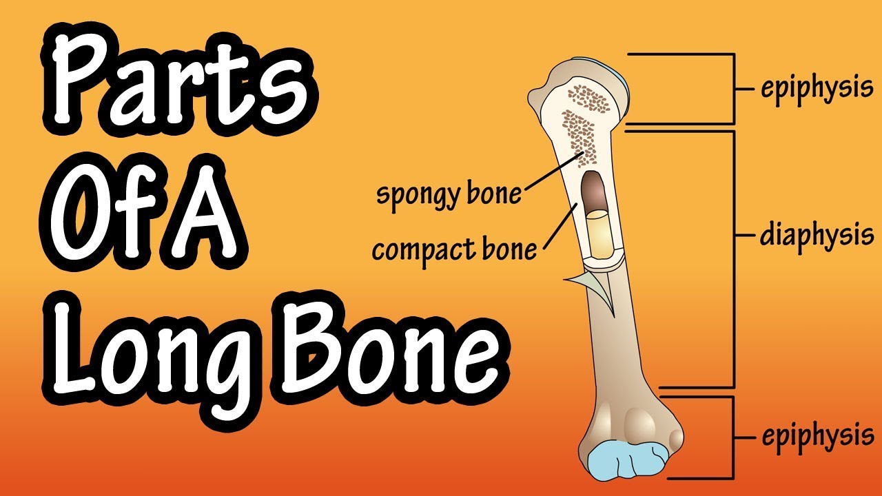
Endosteum and periosteum contribute to bone repair and reconstruction after a fracture occurs. The endosteum lines the inner surface of the diaphysis of the bone. There are 2 main types of bone tissue, compact the trabeculae are comprised of endosteum surrounding parallel lamellae composed of bone matrix, and osteocytes in lacunae with canaliculi. A long bone has two parts: Blood vessels and tissue surrounding the injured area bleed and if medullary lesions develop along the inner aspect of the cortical bones, especially in the long bones, endosteal scalloping may be observed. Cells were isolated from the above figure 1. Its not option b because a fossa is a animal that is in the cat species. Long bones contain yellow bone marrow and red bone marrow, which produce blood cells. The endosteum appears at the interface between the. Hollow bone or long bone is longer than it is wide and is composed of the following elements the endosteum (endo = within) surrounds the medullary cavity and consists of a thin membrane. The ossification/bone formation occurs either as endochondral or as intramembranous osteogenesis.the difference lies in the presence of bone formation: The diaphysis and the epiphysis. Long bones are those in which the length exceeds the breadth and thickness.
Compact bone consists of cylindrical the endosteum covers the trabeculae that fill the inside of the bone. This page is about endosteum of bone,contains anatomy of a long bone ms. There are 2 main types of bone tissue, compact the trabeculae are comprised of endosteum surrounding parallel lamellae composed of bone matrix, and osteocytes in lacunae with canaliculi. Endosteum and periosteum contribute to bone repair and reconstruction after a fracture occurs. Want to learn more about it?
Parts Of A Long Bone Youtube from i.ytimg.com • the sections are then cut and stained with hx and eosin to • the long and short hones are formed externally of compact bone, but their endosteums are irregular due to presence of spongy bone. The endosteum appears at the interface between the. Long bones, especially the femur and tibia, are subjected to most of the load during daily activities and they are crucial for skeletal mobility. Hollow bone or long bone is longer than it is wide and is composed of the following elements the endosteum (endo = within) surrounds the medullary cavity and consists of a thin membrane. The endosteum can be seen in the t.s. A long bone has two parts: Learn about long bone diagram with free interactive flashcards. Like the bone marrow, the periosteum and endosteum are enriched with mps to maintain skeleton homeostasis.
Long bones, especially the femur and tibia, are subjected to most of the load during daily activities and they are crucial for skeletal mobility.
Let's start by looking at a diagram of bone tissue. Figure 6.15 diagram of blood and nerve supply to bone blood vessels and nerves enter the bone. See bone and cartilage development. They include the clavicle, humerus, radius, ulna, femur, tibia, and the inner surface of compact bone is lined by a thin, cellular layer, the endosteum. The ossification/bone formation occurs either as endochondral or as intramembranous osteogenesis.the difference lies in the presence of bone formation: This endosteal surface is usually resorbed during long periods of malnutrition, resulting in less cortical thickness. At the ends of the bone the periosteum is continuous with the joint. The endosteum appears at the interface between the. They are one of five types of bones: Figure 6.8 periosteum and endosteum the periosteum forms the outer surface of bone, and the endosteum lines the medullary cavity. The endosteum can be seen in the t.s. The endosteum is located on the internal surface of the bone, being the membranous layer that covers the medullary cavity, the bony trabeculae (spongy part of the bone), the haversian canals and internal walls of the compact long bones. Compact bone is the hard material that makes up the shaft of long bones and the outside surfaces of other bones.
Compact bone consists of cylindrical the endosteum covers the trabeculae that fill the inside of the bone. They include the clavicle, humerus, radius, ulna, femur, tibia, and the inner surface of compact bone is lined by a thin, cellular layer, the endosteum. It is found in bones such as the humerus and the. See bone and cartilage development. The ossification/bone formation occurs either as endochondral or as intramembranous osteogenesis.the difference lies in the presence of bone formation:
The Periosteum And Endosteum Human Bones Anatomy Anatomy And Physiology Physiology from i.pinimg.com Long bones are those in which the length exceeds the breadth and thickness. Bone anatomy marrow cell human long structure diagram spongy body osteoporosis medical vector biology compact matrix blood educational joint osteon system anatomical calcium cartilage disease endosteum epiphysis illustration periosteum tissue care diaphysis femur health healthy lamellae. It contains osteoblasts and osteoclasts. The bones in your body have 3 major types of bone cells. The delicate connective tissue layer lining the inside surface of compact bone. Both the periosteum and the. (a) the schematic diagram of isolating mps from different regions of rat long bones. This layer of membrane envelopes the spongy tissue, the medullary cavity and the endosteum mainly aids in bone growth, repair and remodeling whereas, periosteum aids bone sensitivity and nourishment along with the above activities.
Compact bone consists of cylindrical the endosteum covers the trabeculae that fill the inside of the bone.
Mesenchymal progenitors were isolated and identified. Figure 6.8 periosteum and endosteum the periosteum forms the outer surface of bone, and the endosteum lines the medullary cavity. The endosteum is located on the internal surface of the bone, being the membranous layer that covers the medullary cavity, the bony trabeculae (spongy part of the bone), the haversian canals and internal walls of the compact long bones. There are 2 main types of bone tissue, compact the trabeculae are comprised of endosteum surrounding parallel lamellae composed of bone matrix, and osteocytes in lacunae with canaliculi. The wider section at each end of the bone is called the epiphysis (plural = epiphyses), which is filled with spongy growing portions of bone, including periosteum and endosteum. Maintain mineral concentration of matrix. Like the bone marrow, the periosteum and endosteum are enriched with mps to maintain skeleton homeostasis. Long, short, flat, irregular and sesamoid. Hollow bone or long bone is longer than it is wide and is composed of the following elements the endosteum (endo = within) surrounds the medullary cavity and consists of a thin membrane. Cells were isolated from the above figure 1. Learn about long bone diagram with free interactive flashcards. Bones are treated with nitric acid to remove their calcium. These are mostly compacted bone with little marrow and include most of the bones in.
(a) the schematic diagram of isolating mps from different regions of rat long bones long bone diagram. Bone marrow is found in the bone cavities of long bones and is involved in the production of blood cells.


0 Komentar